Inside the Faculty of Biological Sciences research facilities
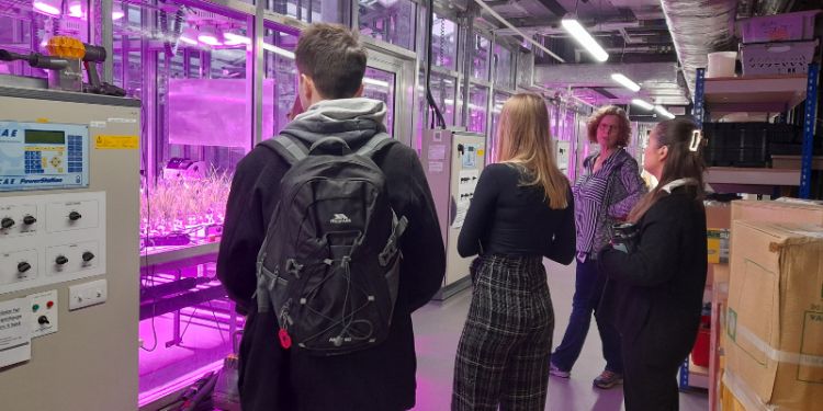
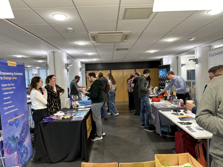
The Faculty of Biological Sciences welcomed colleagues across the University of Leeds to tour its state-of-the-art research facilities on 16 November.
The open day, which was attended by researchers across a range of disciplines, was an opportunity to experience the cutting-edge technology on campus and network with colleagues.
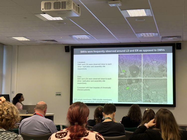
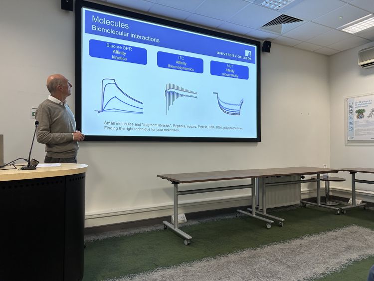
The day encompassed a series of talks and visits to 7 core facilities by facility experts, as well as presentations from previous users.
Some of the innovative facilities on show were the Electron Microscopy facility, a resource which can produce high-resolution structures of macromolecular complexes; Mass Spectrometry, which enables mass spectrometry technologies across a wide range of scientific disciplines, and the Bioimaging and Flow Cytometry facility.
Together they provide a full pipeline for preparation and complete characterisation of systems from single molecules to macro-molecular complexes and cells, revealing the molecular level of cellular function.
Kevin Corke, Director of Operations in the Faculty of Biological Sciences said:
Our cutting-edge facilities here at Leeds are already enabling pioneering research across a range of disciplines thanks to our ongoing investments, such as our newly refurbished lab and office space in Garstang, and the huge pool of talent and expertise we have within the faculty.
“Open days like these not only showcase our cutting-edge equipment but they also bring people together to network and share knowledge, which is a key part of our research culture.”
Dr Sally Boxall, Facility Manager in the Faculty of Biological Sciences added:
Our facilities are available to researchers from across the University, as well as external users from academia and industry. We provide full training and support to ensure that researchers of all levels of expertise and experience can gain the maximum potential from our systems to take their research to the next level.
“Do reach out to our friendly team of experts to discuss your research. There could be a wealth of opportunities waiting to be discovered.”
Upasana Sykora
Dr Iain Manfield
Astbury Biostructure Laboratory Electron Microscopy Facility
The Electron Microscopy facility can generate high-resolution data to produce structures of a broad range of macromolecular complexes including membrane proteins, ribosomes and viruses.
In addition, we have a suite of equipment for the preparation of cells, tissues and organisms.
Bioimaging and Flow Cytometry
The Bioimaging and Flow Cytometry Facility provides access to a range of state-of-the-art light microscopy and flow cytometry technology to support cutting-edge research.
These range from widefield fluorescent systems and confocal microscopes through to super-resolution systems.
We have the capability to image both fixed and live samples to facilitate the investigation of a wide range of biological questions.
The facility also has flow cytometry capabilities as well as a cell sorter to identify and isolate populations of interest.
Biomolecular Mass Spectrometry (MS)
The Biomolecular Mass Spectrometry Facility is a state-of-the-art centre offering service analyses and research opportunities for investigating the mass, structure and conformation of biomolecules and their complexes.
The MS Facility provides access to instrumentation for performing a range of experiments from routine molecular mass measurements and protein identification to more advanced structural proteomics analyses including native MS, hydrogen-deuterium exchange MS and chemical cross linking.
Nuclear Magnetic Resonance (NMR)
NMR has unique capabilities to study systems directly in solution, investigate dynamics, provide atomic resolution data on intrinsically unfolded, partially folded and transiently interacting biological macromolecules, and to screen for small molecule ligands.
Protein Production, Interactions, X-ray and Circular Dichroism (PIXC)
We have a range of expertise in protein expression, protein folding, tissue culture, interactions, and circular dichroism.
Protein production provides a service from Design of Experiment, through cloning, mutagenesis, small-scale expression screening, large-scale growth and high-quality purification.
Circular dichroism spectroscopy is a fast, quantitative and non-destructive photometric technique that can help answer an array of common questions about the stability, structure and interactions of nucleic acids and proteins.
Refeyn mass photometry or Fida flow diffusion can also determine solution molecular weights and hydrodynamics.
More information about our research facilities can be found online.


