What worms can tell us about brains and behaviour
The University of Leeds has collaborated with a leading American institution on research that helps to crack the code that relates brain and behaviour.
Researchers at Massachusetts Institute of Technology (MIT) mapped how neurons across the tiny brain of the microscopic C. elegans worm encode its behaviours – such as movement and feeding – revealing many new insights about its nervous system.
Published in the prestigious journal Cell, the team produced an atlas of how each brain cell encodes the animal’s actions, revealing the underlying “logic” of how the worm’s brain produces a sophisticated and flexible repertoire of behaviours.
Previous pioneering research, carried out by University of Leeds PhD student Elpiniki Kalogeropoulou, from the Schools of Computing and Biology, had genetically engineered a unique set of these creatures to lack a particular set of brain cells – those that affect their ability to turn.
The MIT academics used these distinctive animals to test if their map of the brain’s turning and steering neurons was correct.
The research revealed that when these cells are eliminated, the logic of the brain and its activity changes – exactly as predicted.
Worm bank
Professor Netta Cohen, a Systems and Computational Neuroscientist in the School of Computing at Leeds, co-supervised the PhD research along with Professor Ian Hope, Emeritus Professor from the School of Biology at Leeds.
She assisted with the MIT study and said: “This collaboration demonstrates one of the great advantages of the C. elegans field - that we can easily send specimen from one lab to another – or order them from a ‘worm bank’. It makes it really easy to collaborate and push the science forward.”
The advantage of our worms is that we targeted cells that nobody else had looked at before – and these cells have a very specific effect on turning behaviours, without messing other things up. These worms enabled MIT to test their theories.
Understanding the full relationship between brain activity and behaviour has long proved an insurmountable challenge. But after inventing new technologies and methods for the purpose, a team of scientists in The Picower Institute for Learning and Memory at MIT has produced a rigorous accounting of most neurons in the tiny brain of a humble C. elegans worm.
WormWideWeb
“This study provides a global map of how the animal’s nervous system is organized to control behaviour,” said senior author Steven Flavell, Associate Professor in MIT’s Department of Brain and Cognitive Sciences.
MIT Graduate students Jungsoo Kim and Adam Atanas, who each earned their PhDs this spring for the research, are the study’s co-lead authors.
They have made all their data, and the findings of their model and atlas, freely available to fellow researchers at a website called the WormWideWeb.
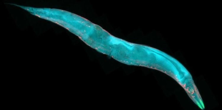
C. elegans are nematodes that feed on bacteria found in rotting vegetation in gardens. They are only around a millimetre in length and as thin as a human hair.
An adult worm has exactly 302 cells in its nervous system - by comparison, the human brain has around 100 billion cells.
But almost two-thirds of the worm’s nerve cells form a ring in the head region, where they make thousands of connections with each other.
This ‘brain’ is the control centre of the animal, where much of the sensing and decision-making takes place. It’s the only animal brain which has been anatomically mapped, so all the cells are known, as well as their neighbours, and how they wire up.
Further Information
“Brain-wide representations of behavior spanning multiple timescales and states in C. elegans” was published in Cell on August 21, 2023.


