Sport and Exercise Sciences Facilities
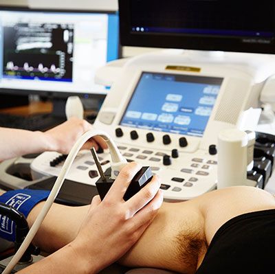
Cardiac and Vascular Imaging Laboratory
Cardiac and Vascular Imaging Laboratory
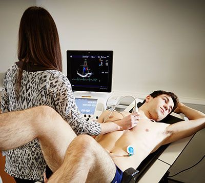
We use ultrasound to explore the structure and function of the heart and major arteries in a variety of different populations (eg healthy aged, heart failure), and to investigate the influence of physiologic/environmental factors (eg exercise, elevated work of breathing, hypoxia) on cardiac and vascular outcomes.
For example, we use Doppler ultrasound to assess brachial and femoral artery blood flow during recumbent cycle exercise, and evaluate how changes in exercise-induce blood flow alters vascular function (via flow mediated dilatation).
We also use ultrasound to characterise arterial wall thickness, stiffness, compliance and distensisibility. The use of ultrasound also allows us to extensively assess left ventricular structure and function, as well as obtain estimates of pulmonary arterial pressure and pulmonary vascular resistance.
Through our links with collaborators at Leeds General Infirmary, we can also measure aortic blood flow and cardiac perfusion at rest and during exercise via cardiac MRI with a specialised cycle ergometer.
Key equipment: GE Vivid E9 ultrasound; GE Portable Vivid iq ultrasound; Vascular Imager data acquisition system; Brachial and Carotid Tools data analysis system, LODE Angio cycle ergometer for recumbent or supine cycling; SphygmaCor tonometer.
Contacts: Prof Karen Birch (k.m.birch@leeds.ac.uk).